Compliance
Quality Management System
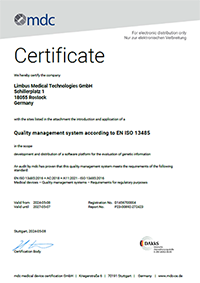
varvis® is developed under the EN ISO 13845 certified quality management system. The standard ensures that the medical device consistently meets customer expectations of quality, safety, and performance.
Versioning Scheme
Version numbers of the software including its sub-modules follow semantic versioning, i.e. a numeric schema major.minor.patch and the following semantics:
-
Major versions reflect substantial changes *; i.e. changes that have impact in terms of the intended purpose of the device or the device performance and/or safety
-
Minor versions reflect non-substantial changes in the features and functionality of the software
-
Patch versions reflect bug fixes and maintenance releases
* 'Substantial changes' refer to paragraph 2.4, Annex IX, IVDR
Safety and performance information
In the following, performance and safety information is provided for the entire device, including its variants P001-01and P001-02.
The confusion matrix of statistical classification is shown below with the abbreviations used in this document.
|
|
Comparator method |
Total |
|||
|
Positive |
Negative |
||||
|
Test |
Positive |
TP |
FP |
TP+FP |
|
|
Negative |
FN |
TN |
FN+TN |
||
|
No calls or invalid calls |
E |
F |
E+F |
||
|
|
Total |
TP+FN+E |
FP+TN+F |
N |
|
Please note that not all entries in the confusion matrix can be determined for all genomic variant types [1]. For example, theoretically, there are an unlimited number of Indel variants, which means that TN cannot be determined for SNV/Indel discovery. The performance characteristics that were determined for this device are:
|
Performance characteristic |
Abbr. |
Formula |
Variant type |
Alias |
|---|---|---|---|---|
|
Positive Percent Agreement |
PPA |
TP/(TP+FN) |
SNV/Indel, CNV |
Sensitivity |
|
Negative Percent Agreement |
NPA |
TN/(TN + FP) |
CNV |
Specificity |
|
Technical Positive Predictive Value |
TPPV |
TP/(TP + FP) |
SNV/Indel, CNV |
Precision |
|
Limit of Detection |
LoD |
- |
SNV/Indel, somatic |
- |
|
Limit of Blank |
LoB |
- |
SNV/Indel, somatic |
- |
|
Reproducibility and Repeatability |
- |
- |
SNV/Indel |
- |
Detection of SNV/Indels in NGS data (germline)
The analytical performance parameters for SNV and Indel discovery in germline samples are listed below (in %) in conjunction with the confidence intervals.
|
Parameter |
Variant type |
WGS |
WES |
NGS panel |
|
PPA |
SNV |
99.8 |
99.4 |
99.1 |
|
PPA 95% CI |
SNV |
99.8..99.8 |
99.4..99.4 |
98.9..99.3 |
|
TPPV |
SNV |
99.8 |
99.5 |
99.8 |
|
TPPV 95% CI |
SNV |
99.8..99.8 |
99.5..99.6 |
99.7..99.9 |
|
PPA |
Indel |
97.1 |
86.9 |
71.9 |
|
PPA 95% CI |
Indel |
97.1..97.2 |
86.7..87.1 |
69.3..74.4 |
|
TPPV |
Indel |
95.9 |
72.4 |
56.9 |
|
TPPV 95% CI |
Indel |
95.9..96.0 |
72.1..72.7 |
54.4..59.4 |
|
PPA |
all |
99.5 |
98.3 |
96.7 |
|
PPA 95% CI |
all |
99.4..99.5 |
98.3..98.3 |
96.4..97.0 |
|
TPPV |
all |
99.2 |
96.9 |
95.0 |
|
TPPV 95% CI |
all |
99.2..99.2 |
96.8..96.9 |
94.6..95.4 |
Confidence intervals were calculated using the Wilson score method [2].
The performance was achieved with data sets of the following quality:
|
Analyte |
Depth of coverage |
% bases covered at 20x |
Insert size |
On-target rate |
|---|---|---|---|---|
|
WGS |
> 90 |
n/a |
> 300 |
n/a |
|
WES |
> 80 |
> 96% |
> 180 |
> 75% |
|
NGS panel |
> 320 |
> 99% |
> 200 |
> 75% |
Note that the analytical performance of NGS-based tests strongly depends on quality parameters like the depth of coverage and the selected target region [3].
It is therefore recommended to determine the performance characteristics for the particular NGS assay and laboratory process according to applicable guidelines [4]–[7].
Reproducibility and repeatability
Repeatedly processing the same data set with the device yields identical results (100% repeatability). Reproducibility was assessed using replicates of the same DNA sample thereby keeping conditions like the type of sequencing instrument, the day of measurement, the operating conditions remain the same. This means that only the sequencing process of the DNA is repeated which represents a sampling of different NGS reads.
This assessment of reproducibility therefore encompasses
-
Variation in the preparation of DNA replicates (e. g. amount of DNA),
-
Variation due to errors in the sequencing process,
-
Variation due to different local optimization of alignment in each sampling.
Under these conditions the PPA varied by 0.2% and the TPPV varied by 0.3%.
Limitations
The following limitations should be noted:
-
Genome-in-a-bottle samples have been characterized using NGS technologies, i. e. that the reference data set is biased towards variants that can be easily discovered by NGS.
-
In addition, the performance evaluation is restricted to the trusted regions of the GiaB data sets. Typically, these trusted regions represent those genomic regions that can easily be assessed using NGS.
-
The analytical performance outside trusted regions should be expected to be lower than reported here [3].
-
The performance evaluation was performed within the manufacturer’s target regions plus 50bp around these regions. This is the region that is typically evaluated in practice. The performance reported here may be lower than when reporting limited to the manufacturer’s target regions.
Detection of SNV/Indels in NGS data (somatic)
The performance characteristics of the somatic analysis are provided in % in the table below.
|
Parameter |
Variant type |
WGS |
|---|---|---|
|
Genome build |
|
hg38 |
|
Limit of blank |
|
10.0 |
|
Limit of detection |
|
15.0 |
|
PPA |
SNV |
97.7 |
|
PPA 95% CI |
SNV |
97.7..97.8 |
|
TPPV |
SNV |
99.3 |
|
TPPV 95% CI |
SNV |
99.2..99.3 |
|
PPA |
Indel |
79.0 |
|
PPA 95% CI |
Indel |
78.6..79.5 |
|
TPPV |
Indel |
93.8 |
|
TPPV 95% CI |
Indel |
93.5..94.0 |
|
PPA |
all |
95.6 |
|
PPA 95% CI |
all |
95.5..95.7 |
|
TPPV |
all |
98.7 |
|
TPPV 95% CI |
all |
98.7..98.8 |
These performance characteristics include both germline and somatic variants. The WGS data set, a mixture of two GiaB samples, had an average depth of coverage of 110.
Detection of CNV in NGS data
General
The analytical performance does not depend on the genome build. hg38 or GRCh37 provide equivalent performance.
The CNV discovery of this device presumes that CNVs and pathogenic CNVs in particular are rare. CNVs that are present in 1 out of a pool of 5 samples or in 4 out of a pool of 10 can be detected with high sensitivity. This must be considered when analyzing data from families. CNVs with high prevalence or polymorphisms cannot reliably be detected with the device.
Since the performance characteristics of a specific NGS assay depend on the configuration of the assay and the laboratory process (e. g. sequencing capacity, depth of coverage, uniformity across samples), it is recommended to perform an assay-specific validation according to applicable guidelines [4]–[7].
Performance evaluation using public patient data
A performance assessment using the public data set ICR96 [8] comprising 96 patient samples is provided here. The data set is publicly available and may serve as a benchmark between different devices.
Note that the ICR96 data set has been characterized as “less homogeneous” than other data sets by the reviewers of the publication. It should be expected that a results with a more modern laboratory set up can generate data of greater uniformity and therefore better analytical performance. The results reported here therefore represent a lower limit.
The quality thresholds are:
|
Parameter |
Description |
Threshold |
|
Reference spread (RefSpread) |
Quality value assigned to a single target region |
< 0.2 |
|
Bivariance (bivar) |
Quality value assigned to a data set as a whole |
< 0.2 |
The following table summarizes the performance characteristics from the ICR96 data set:
|
Name |
Loss |
Gain |
All |
|
PPA |
98.8% |
97.5% |
98.4% |
|
PPA 95% CI |
98.82..98.82% |
97.53..97.53% |
98.40..98.40% |
|
NPA |
99.9% |
100.0% |
99.8% |
|
NPA 95% CI |
99.86..99.86% |
99.96..99.96% |
99.82..99.82% |
The following table indicates how many samples and how many individual target regions were excluded from the analysis (no-calls) after application of the quality thresholds:
|
Parameter |
Result |
Fraction |
|
Total number of samples |
96 |
100.0% |
|
Samples failing thresholds |
10 |
10.4% |
|
Total number of targets |
31203 |
100.0% |
|
Targets failing thresholds |
417 |
1.3% |
The choice of reference genome (hg38 vs GRCh37) has no significant impact on the overall analytical performance regarding the data set. It should be noted, though, that the analysis using hg38 enhances the mapability of NGS reads and leads to a larger number of samples being acceptable for analysis. Partial deletions or mosaic variations were not assessed.
Detection of STRs in NGS data
The analytical performance of detecting STRs ≥ 5bp based on PacBio HiFi long-read whole-genome sequencing data for the GiaB reference sample HG002 is detailed below. For each performance characteristic, point estimates, the lower bound of their 95% confidence intervals (calculated using the Wilson score method), and the acceptance criteria are presented.
|
Performance characteristic |
Acceptance criterion |
Analytical result |
|
TPPV / % |
>=80 |
94.3 |
|
TPPV 95% CI Lower Bound / % |
includes or exceeds 80 |
94.1 |
|
PPA / % |
>=85 |
96.5 |
|
PPA 95% CI Lower Bound / % |
includes or exceeds 85 |
96.4 |
|
NPA / % |
>=98 |
99.5 |
|
NPA 95% CI Lower Bound / % |
includes or exceeds 98 |
99.5 |
|
NPV / % |
>=98 |
99.7 |
|
NPV 95% CI Lower Bound / % |
includes or exceeds 98 |
99.7 |
It is notable that the long-read STR analysis of the medical device is conducted for the GRCh38 human reference genome.
Overall, the analytical performance of the medical device’s STR analysis is high and acceptable across the entire genome. A stratified analysis supports this strong result, confirming the STR analysis’s consistently high and acceptable performance across various expected total repeat lengths. Importantly, the medical device also demonstrates superior STR calling performance compared to GangSTR, a common short-read method used for STR analysis.
Since the performance analysis covers such an extensive set of repeat regions, the findings are highly applicable to calling STR sequences with a minimum total length of 5bp across the entire genome, even acknowledging that specific genomic contexts may present unique challenges.
Detection of SVs in NGS data
The analytical performance of detecting SVs ≥ 50bp based on PacBio HiFi long-read whole-genome sequencing data for the GiaB reference sample HG002 is detailed below. For each performance characteristic, point estimates, the lower bound of their 95% confidence intervals (calculated using the Wilson score method), and the acceptance criteria are presented.
|
Performance characteristic |
Acceptance criterion |
Analytical result |
|
PPA / % |
>=86 |
95.8 |
|
PPA 95% CI Lower Bound / % |
includes or exceeds 86 |
95.5..96.1 |
|
TPPV / % |
>=85 |
99.0 |
|
TPPV 95% CI Lower Bound / % |
includes or exceeds 85 |
98.9..99.2 |
It is notable that the long-read SV analysis of the medical device is conducted for the GRCh38 human reference genome.
Overall, the analytical performance of the medical device’s SV analysis is high and acceptable across the entire genome. While the analysis concentrates on insertions and deletions with a minimum length of 50bp as specific SV subtypes, its extensive genome-wide coverage of SVs ensures that the performance findings are broadly applicable to SV calling. This holds true even when acknowledging that specific genomic contexts might present unique challenges. A stratified analysis further supports this strong result, confirming consistently high and acceptable performance for deletions of any size, and for insertions up to 5000bp.
References
[1] P. Krusche et al., “Best practices for benchmarking germline small-variant calls in human genomes,” Nat Biotechnol, vol. 37, no. 5, Art. no. 5, May 2019, doi: 10.1038/s41587-019-0054-x.
[2] R. G. Newcombe, “Improved confidence intervals for the difference between binomial proportions based on paired data,” Statistics in Medicine, vol. 17, no. 22, pp. 2635–2650, Nov. 1998, doi: 10.1002/(SICI)1097-0258(19981130)17:22<2635::AID-SIM954>3.0.CO;2-C.
[3] M. H. Cleveland, J. M. Zook, M. Salit, and P. M. Vallone, “Determining Performance Metrics for Targeted Next-Generation Sequencing Panels Using Reference Materials,” The Journal of Molecular Diagnostics, vol. 20, no. 5, pp. 583–590, Sep. 2018, doi: https://doi.org/10.1016/j.jmoldx.2018.04.005.
[4] C. Rehder et al., “Next-generation sequencing for constitutional variants in the clinical laboratory, 2021 revision: a technical standard of the American College of Medical Genetics and Genomics (ACMG),” Genetics in Medicine, pp. 1–17, Apr. 2021, doi: 10.1038/s41436-021-01139-4.
[5] E. Souche et al., “Recommendations for whole genome sequencing in diagnostics for rare diseases,” Eur J Hum Genet, pp. 1–5, May 2022, doi: 10.1038/s41431-022-01113-x.
[6] G. Matthijs et al., “Guidelines for diagnostic next-generation sequencing,” Eur J Hum Genet, Oktober 2015, doi: 10.1038/ejhg.2015.226.
[7] P. Bauer, “S1 Leitlinie: Molekulargenetische Diagnostik mit Hochdurchsatz-Verfahren der Keimbahn, beispielsweise mit Next-Generation Sequencing,” medgen, vol. 30, no. 2, pp. 278–292, Jun. 2018, doi: 10.1007/s11825-018-0189-z.
[8] S. Mahamdallie et al., “The ICR96 exon CNV validation series: a resource for orthogonal assessment of exon CNV calling in NGS data,” Wellcome Open Res, vol. 2, p. 35, May 2017, doi: 10.12688/wellcomeopenres.11689.1.
